
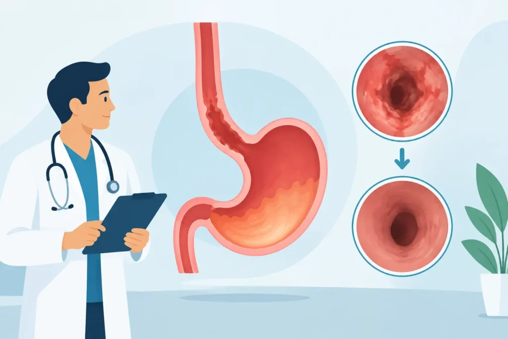
A Barrett’s esophagus diagnosis can feel alarming, especially when the conversation turns to cancer risk. The encouraging news is that Barrett’s esophagus treatment outcomes are often very favorable when care is based on the precise cell changes seen on biopsy, the extent of disease, reflux control, and consistent follow-up with a gastroenterology team.
Barrett’s esophagus occurs when long-term acid reflux changes the lining of the lower esophagus. Most people with Barrett’s esophagus will not develop esophageal cancer. Still, the condition deserves thoughtful surveillance and, when dysplasia is present, timely treatment to remove or destroy abnormal tissue before it can progress.
What Determines Barrett’s Esophagus Treatment Outcomes?
The most significant factor is whether biopsies show dysplasia, meaning precancerous changes in the Barrett’s cells. Pathologists generally classify these findings as no dysplasia, indefinite for dysplasia, low-grade dysplasia, or high-grade dysplasia. Because the diagnosis guides major treatment decisions, a finding of dysplasia is often reviewed by an experienced gastrointestinal pathologist.
For patients without dysplasia, the main goal is reducing reflux exposure and monitoring the esophagus at appropriate intervals. For patients with confirmed low-grade or high-grade dysplasia, the goal shifts to eliminating the abnormal Barrett’s lining through endoscopic therapy. When treatment is recommended at the right stage, it can substantially lower the chance that advanced precancerous tissue will progress to cancer.
Outcomes also depend on several personal factors: the length of the Barrett’s segment, whether visible nodules are present, the quality of acid control, smoking status, body weight, family history, and whether follow-up care is completed. A plan should account for the full clinical picture rather than relying on one biopsy result alone.
Expected Outcomes by Barrett’s Esophagus Stage
Barrett’s esophagus without dysplasia
If biopsies show no dysplasia, many patients do not need an ablative procedure. Proton pump inhibitor medications are commonly used to reduce acid exposure, improve reflux symptoms, and support healing of inflammation. Your physician may also discuss practical reflux measures, such as avoiding late meals, identifying food triggers, elevating the head of the bed when nighttime reflux is a concern, and addressing excess weight when appropriate.
Surveillance upper endoscopy remains a key part of care. During the procedure, the gastroenterologist closely examines the esophageal lining and takes systematic biopsies to look for changes that may not be visible to the eye. The interval between endoscopies varies based on the length of Barrett’s tissue and individual risk factors, but it is typically measured in years rather than months when no dysplasia is present.
The outcome patients most often want to hear is that the cancer risk remains low for nondysplastic Barrett’s esophagus. Surveillance is not passive care. It is a preventive strategy designed to identify change early, when treatment is most effective.
Indefinite for dysplasia
Inflammation from uncontrolled reflux can make cells difficult to interpret. If pathology is reported as indefinite for dysplasia, the next step is often to optimize acid-suppressing treatment and repeat endoscopy after the tissue has had time to heal. This approach helps distinguish inflammation-related atypia from true precancerous change.
This category can be frustrating because it does not always produce an immediate answer. However, reassessment prevents patients from being overtreated for uncertain findings while ensuring that meaningful dysplasia is not overlooked.
Low-grade dysplasia
Confirmed low-grade dysplasia carries a higher progression risk than nondysplastic Barrett’s esophagus. Endoscopic eradication therapy is frequently recommended, particularly when expert pathology review confirms the diagnosis. The most common approach combines removal of any raised or suspicious areas with treatment that destroys the remaining flat Barrett’s tissue.
Radiofrequency ablation uses controlled heat to treat the superficial lining of the esophagus. In selected patients, cryotherapy, which uses extreme cold to destroy abnormal tissue, may also be considered. These procedures are performed through an endoscope, not through open surgery.
For many patients, treatment can eliminate dysplasia and achieve complete eradication of intestinal metaplasia, the tissue change associated with Barrett’s esophagus. Multiple treatment sessions may be necessary, especially when the Barrett’s segment is longer. Temporary chest discomfort, painful swallowing, or narrowing of the esophagus can occur, but serious complications are uncommon when therapy is performed by an experienced endoscopy team.
High-grade dysplasia or early esophageal cancer
High-grade dysplasia and very early cancer require prompt evaluation, but they do not automatically mean that major surgery is needed. When disease is confined to the superficial layers of the esophagus, endoscopic mucosal resection can remove visible nodules or early cancer areas for precise diagnosis and treatment. The remaining Barrett’s tissue is then typically treated with ablation.
For appropriately selected patients, endoscopic therapy offers excellent organ-preserving outcomes and can avoid esophagectomy, the surgical removal of part or all of the esophagus. Surgery, chemotherapy, or radiation may still be needed if cancer has invaded more deeply or spread beyond the surface layers. That is why careful staging and expert evaluation matter from the beginning.
Why Follow-Up Matters After Successful Treatment
A clear endoscopy after ablation is an important milestone, but it is not the end of care. Barrett’s tissue and dysplasia can recur, sometimes at the junction where the esophagus meets the stomach. Continued surveillance allows the care team to identify small areas of recurrence before they become a larger concern.
Most patients continue acid-suppressing medication after eradication therapy, unless their physician recommends otherwise. Persistent reflux can contribute to recurrent injury, and some people have reflux even when they do not feel classic heartburn symptoms. Your treatment plan may also include evaluation for a hiatal hernia or discussion of anti-reflux procedures when medication does not adequately control reflux.
Long-term results are strongest when patients keep surveillance appointments, take medications as directed, and report new symptoms promptly. Difficulty swallowing, unintentional weight loss, vomiting blood, black stools, persistent chest pain, or worsening reflux should be evaluated without delay.
Improving Your Individual Outlook
There is no single outcome timeline that applies to every person with Barrett’s esophagus. A short segment without dysplasia has different implications than a long segment with confirmed high-grade dysplasia. Likewise, a patient with severe obesity, fatty liver disease, diabetes, or chronic reflux may benefit from coordinated care that addresses the metabolic and digestive factors affecting overall health.
Weight management can be clinically relevant for some patients because excess abdominal weight may increase pressure on the stomach and worsen reflux. That does not mean weight loss replaces surveillance or endoscopic treatment. It means a physician-guided plan can support reflux control alongside the care of Barrett’s esophagus. Smoking cessation is equally valuable, since smoking is associated with increased risk of progression and can affect healing.
At NJ Gastroenterology & Health Wellness, a Barrett’s esophagus plan begins with a careful review of your endoscopy findings, pathology, reflux symptoms, medications, personal history, and goals. Board-certified gastroenterology care, advanced endoscopic evaluation, and coordinated support for digestive and metabolic health can help make each next step clear.
When to Discuss Treatment With a Gastroenterologist
Schedule a gastroenterology evaluation if you have Barrett’s esophagus and have not had recommended surveillance, if a biopsy has shown dysplasia or an uncertain result, or if reflux symptoms continue despite treatment. Bring prior endoscopy and pathology reports when possible. They help your physician compare findings over time and determine whether observation, medication adjustment, repeat testing, or endoscopic eradication therapy is appropriate.
The most reassuring path is not guessing about risk. It is having a clear, individualized plan that protects the esophagus now and gives you a reliable framework for long-term care.